In medical term, it is called as Occipital neuralgia It is a condition in which the nerves that go from the top of the spinal cord up via the scalp, named the occipital nerves, are swollen or injured And you might feel pain in back of your head at base of your skull and NauseaHuman skeleton, skull front and back The human skull is generally considered to consist of twentytwo bones %u14 eight cranial bones and fourteen facial skeleton bones In the neurocranium these are the occipital bone, two temporal bones, two parietal bones, the sphenoid, ethmoid and frontal bones Axial muscles of the head, neck, and back It offers protection to the brain, eye balls, inner ears, and nasal passages The skull includes the upper jaw and the cranium It supports and protects the face and the brain In order to be light, the skull is made up by flat and irregular bones, and has hollow spaces called the sinuses Learn more

Human Skull With Veins And Arteries Rear View Vascular System Of The Human Head Viewed From The Back Computer 3d Rendering Canstock
Back of head skull anatomy
Back of head skull anatomy-This is the second video about the skull anatomy and the bones of the head https//wwwAnimatedAnatomycom/ Click To Buy Our Anatomical Software And LBack of skull Anatomy Overview, Anterior Skull Base, Middle Skull Base The greater portion of the anterior floor is convex and grooved by the frontal lobe gyri This portion of the skull base consists of the orbital portion of the frontal bone The ethmoid bone forms the central part of the floor, which is the deepest area of


Human Body Skull Anatomy External Occipital Protuberance Human Back Of Skull Face Human Png Pngegg
13,276 anatomy of the head and neck stock photos, vectors, and illustrations are available royaltyfree See anatomy of the head and neck stock video clips of 133 muscle head anatomy vocal organ diagram female neck anatomy neck wireframe head neck human anatomy head artery anatomy face pharynx vector neck degree head anatomy 3dThe facial bones (14 bones 2zygomatic, 2maxillary, 2palatine, 2nasal, 2lacrimal, vomer, 2inferior conchae, mandible) The occipital bone joins with the atlas near the foramen magnum, a large hole ( foramen) at the base of the skull The atlas joins The nerves of the head and neck include the most vital and important organs of the nervous system — the brain and spinal cord — as well as the organs of the special senses In addition, in this region we also find the major cranial and spinal nerves that connect the central nervous system to the organs, skin, and muscles of the head and neck
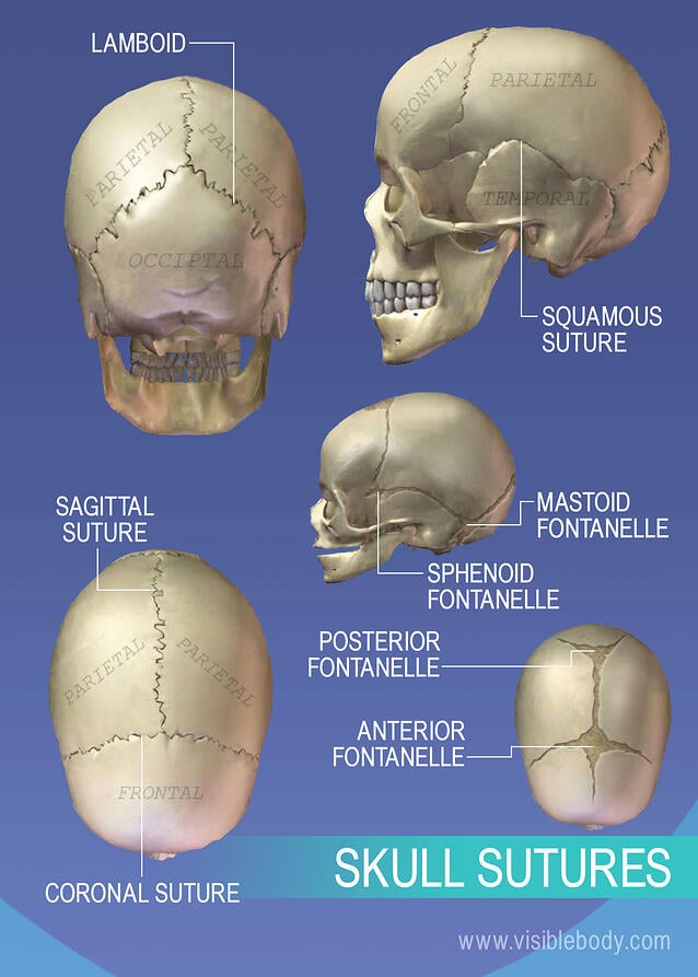
Skull anatomy Frontal Bone Parietal Bone Sagital Suture Coronal Suture the large cranial bone forming the front part of the cranium either of two skull bones between the frontal and occipital bo Located on the midline of the skull lies between both parietal the suture between the parietal and frontal bones of the skullUseful xray images head and neck anatomy stock pictures, royaltyfree photos & images black human anatomy icons head and neck anatomy stock illustrations anatomy, veins of face and neck, sinuses dura mater, diploe, victorian anatomical drawing 19th century head and neck anatomy stock illustrationsThe head is comprised of many bones that form a shelllike bony structure that protects the brain the skull While the bony framework of the neck is define
What is the Pain in Back of Head at Base of Skull ? 10 / 10 ( 1 vote ) External Anatomy Of The Right Side Of The Skull In this image, you will find coronal suture, frontal bone, parietal bone, sphenoid bone, greater wing, squamous suture, lambdoid suture in it You may also find an occipital bone, temporal bone, ethmoid bone, lacrimal bone, lacrimal fossa, zygomatic process, nasal boneBack of head, back part of the head or skull (Anatomy) occiput () Flemish Anatomist who made discoveries about Human Anatomy which surpassed those


Occipital Surgery For Flat Spots On Head Dr Barry L Eppley
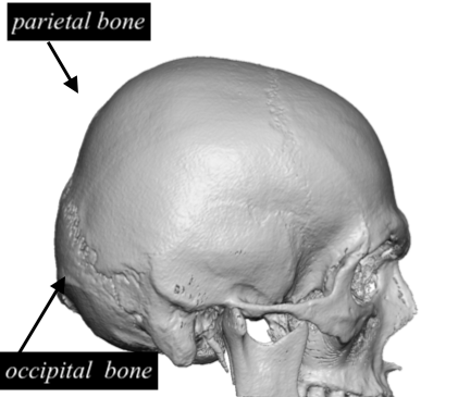


Flat Back Of Head Skull Anatomy Dr Barry Eppley Indianapolis Explore Plastic Surgery
The Organs of the Head include the ear, the eye, the nose and sinuses, the salivary glands, and the oral cavity The ear can be divided in to three sections the external ear, the middle ear, and the inner earThe external ear functions to capture and direct sound waves through the external acoustic meatus to reach the tympanic membrane (ear drum) ) The tympanic membrane marks The cranium (also known as the neurocranium) is formed by the superior aspect of the skull It encloses and protects the brain, meninges, and cerebral vasculature Anatomically, the cranium can be subdivided into a roof and a base Cranial roof – comprised of the frontal, occipital and two parietal bonesEpidural hematoma Bleeding between the tough tissue (dura) lining the inside of the skull and the skull itself, usually shortly after a head injury Initial mild symptoms can progress rapidly to


Neck Anatomy Britannica


Back View Of Human Skeleton Of Head Stock Photo Picture And Royalty Free Image Image
Translation for 'back part of the skull which protrudes outward (Anatomy), bony projection on the back of the head' in English>English dictionary Search over 14 million words and phrases in more than 490 language pairsAbout Skull Anatomy Skull reshaping is done on any of the structures that lie above the face The simplest way to make the difference between the head and the face is to envision a ring that wraps around the head at the level of the eyes Anything above is the head or skull and what lies below is the face This can also be called theThe orbicularis oris is a circular muscle that moves the lips, and the orbicularis oculi is a circular muscle that closes the eye The occipitofrontalis muscle moves up the scalp and eyebrows The muscle has a frontal belly and an occipital (near the occipital bone on the posterior part of the skull) belly In other words, there is a muscle on the forehead (frontalis) and one on the back of


Occipital Bun Wikipedia
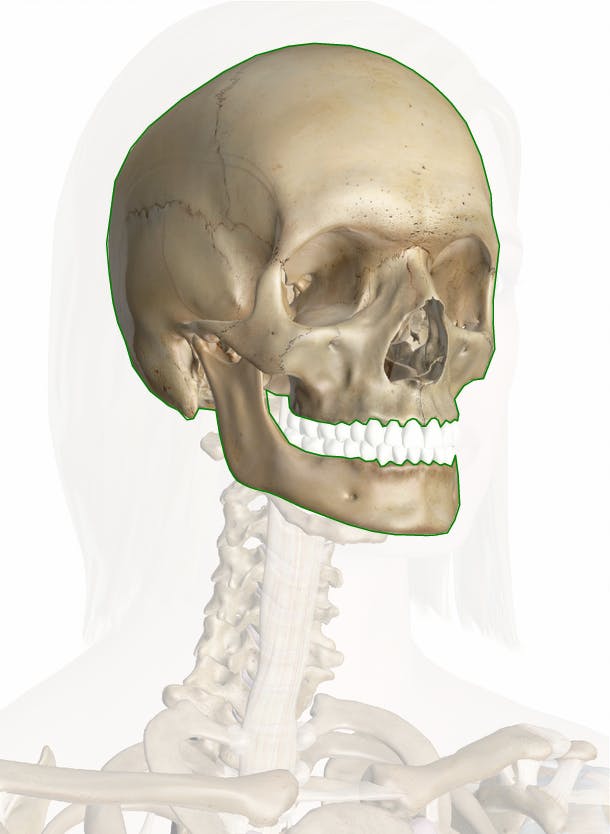


Skull Anatomy Pictures And Information
They move the head in every direction, pulling the skull and jaw towards the shoulders, spine, and scapula Working in pairs on the left and right sides of the body, these muscles control the flexion and extension of the head and neck Working individually, these muscles rotate the head or flex the neck laterally to the left or rightThe cranial base, also known as the base of the skull or skull ba s e, is the most inferior part of the skull forming the floor of the cranial cavity The cranial base is formed by six different bones the ethmoid, sphenoid, occipital, frontal, paired parietal and temporal bones The spine is the backbone of the human skeleton Together, the bones of the skull support the structure of the face and shelter the brain and brain stem The spine provides support to hold the head and body up straight Learn how the housefly uses its anatomy to survive and why housefly meals arrive through a plunger


Human Skull With Veins And Arteries Rear View Vascular System Of The Human Head Viewed From The Back Computer 3d Rendering Canstock
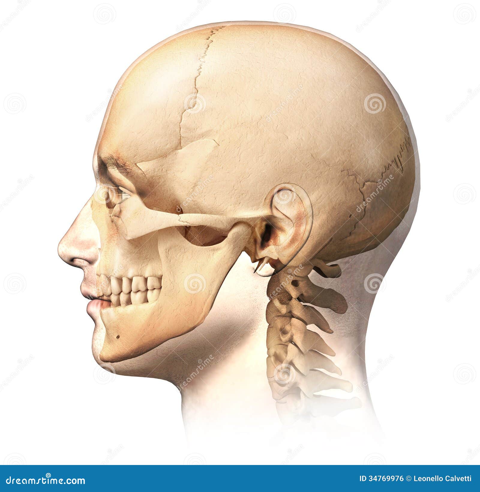


Male Human Head With Skull In Ghost Effect Side View Stock Illustration Illustration Of Inside Human
There are a wide variety of causes of these bumps In addition, each human skull has a natural bump on the back of the head This bump, An anatomy lesson is a good place to start This article will help you understand key anatomical structures in the skull and spine, with the goal of helping you better understand your condition The Craniovertebral Junction Where Your Head and Neck MeetThe occipital bone is the trapezoidshaped bone at the lowerback of the cranium (skull) The occipital bone houses the back part of the brain and is one of seven bones that come together to form the skull It is located next to five of the cranium bones


Upper Cervical Spine Disorders Anatomy Of The Head And Upper Neck


Skull Scalp And Superficial Face
Routine non–contrast material–enhanced head CT is one of the most frequently ordered studies in the emergency department Skull base–related pathologic entities, often depicted on the first or last images of a routine head CT study, can be easily overlooked in the emergency setting if not incorporated in the interpreting radiologist's search pattern, as the3D interactive models and tutorials on the anatomy of the head and face, including the musculature, osseus strutures, ear, orbit, nasal cavity and more!The Crown begins at the point where the top of the head begins to curve downward to the back of the head and ends at the point just above the Occipital bone It is a semicircular area Occipital BoneThe Occipital Bone is the small bony protrusion at
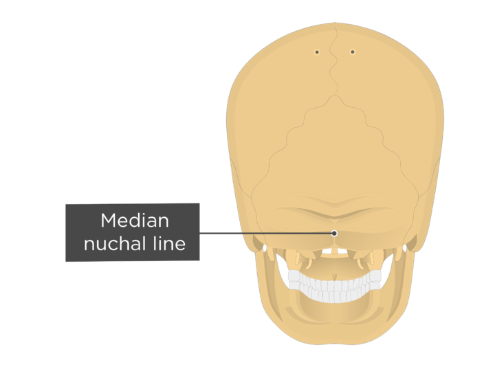


Occipital Bone Anatomy


Axial Skeleton Learn Skeleton Anatomy
Near the base of the skull are a group of muscles called the suboccipital muscles There are 4 paired muscles that attach to the upper two cervical bones (1) The suboccipital muscles can become tense and tender due to factors such as eye strain, wearing new eyeglasses, poor ergonomics at a computer workstation, poor posture, and traumaThe spine provides support to hold the head and body up straight Explore how exactly your eyes help you see the world around you Anatomy is the identification and description of the structures of living things Upper Back Anatomy Sutures of the skull Anatomy Kenhub On this page, you'll find links to descriptions and pictures of theBrowse 4,815 head and neck anatomy stock photos and images available, or search for head and neck cancer or squamous cell carcinoma to find more great stock photos and pictures Houman body parts flat line icons set Man, woman head, brain nose, mouth, foot, ear, lips vector illustration


The Skull Boundless Anatomy And Physiology
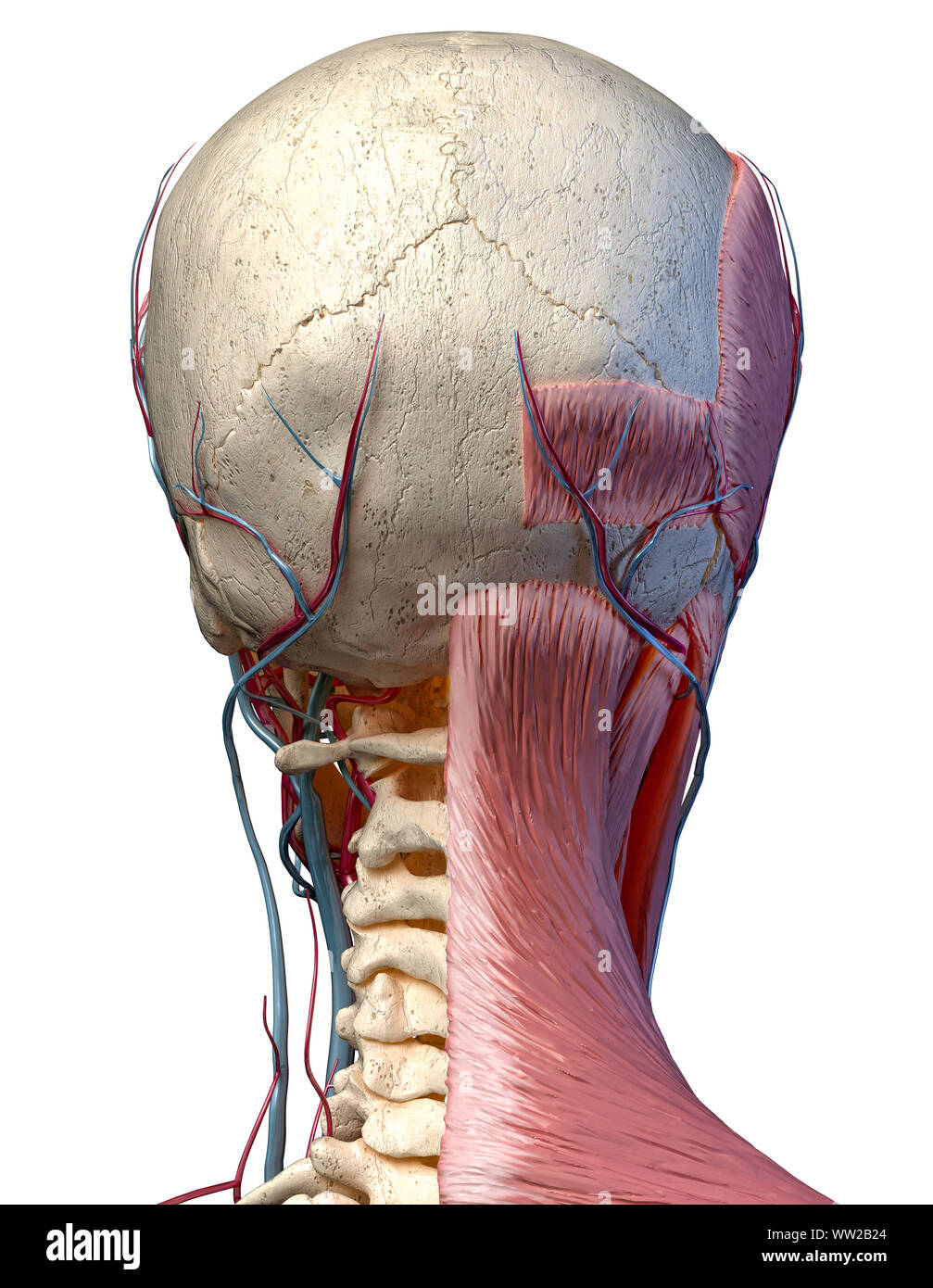


Human Anatomy 3d Illustration Of Head With Skull Blood Vessels And Muscles On White Background Rear View Stock Photo Alamy
115,084 human skull anatomy stock photos, vectors, and illustrations are available royaltyfree See human skull anatomy stock video clips of 1,151 skull system anatomy of skull anatomy skull skull lateral view skull anatomy occipital medical skeleton named facial bone anatomy mandible maxilla human anatomy lateral skull anatomy It is a complex anatomical structure weighing up to five kilograms that rests on the bony skull and in turn, the neck In addition to the evident ears, eyes, nose, and mouth, the head supports a variety of other important structures Muscles of mastication Facial muscles1 Occipital Condyles where the skull articulates with the first cervical vertebrae 2 foramen magnum is also seen, which is where the spinal cord leaves the skull 3 sphenoid sinus This sinus drains via the aperture of the sphenoid sinus (top of body on both sides anteriorly) 4


Bones Of The Skull Learn In 4 Minutes Youtube
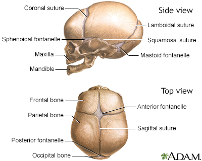


Cranial Sutures Information Mount Sinai New York
The orbicularis oris is a circular muscle that moves the lips, and the orbicularis oculi is a circular muscle that closes the eye The occipitofrontalis muscle moves up the scalp and eyebrows The muscle has a frontal belly and an occipital (near the occipital bone on the posterior part of the skull) belly In other words, there is a muscle on the forehead (frontalis) and one onFlat Back of the Head One of the most common aesthetic deformities of the entire skull is the flat back of the head Well known to occur from sleeping positions in infants and predisposed to occur in certain ethnic groups, lack of a nice shape to the back of The skull base forms the floor of the cranial cavity and separates the brain from other facial structures This anatomic region is complex and poses surgical challenges for otolaryngologists and neurosurgeons alike Working knowledge of the normal and variant anatomy of the skull base is essential for effective surgical treatment of disease in this area
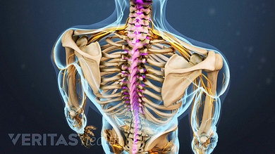


Spinal Anatomy And Back Pain


Massage For Neck Pain Headaches Suboccipitals
The skull base forms the floor of the cranial cavity and separates the brain from other facial structures From an anatomical perspective, the skull is divided into two parts Inferior view of base of the skull Axial muscles of the head, neck, and back Learn skull anatomy with skull bones quizzes and diagram labeling exercises The spine is the backbone of the human skeleton Anatomy is the science that studies the structure of the body The shoulder is designed to be incredibly flexible, enabling a wide range of motion The muscles of the neck run from the base of the skull to the upper back and work together to bend the head and assist in breathing
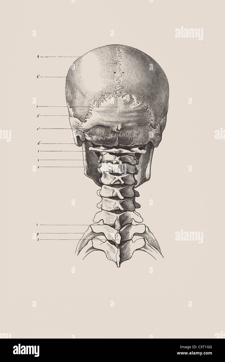


Cranial Skeleton From The Back Anatomical Illustration Stock Photo Alamy
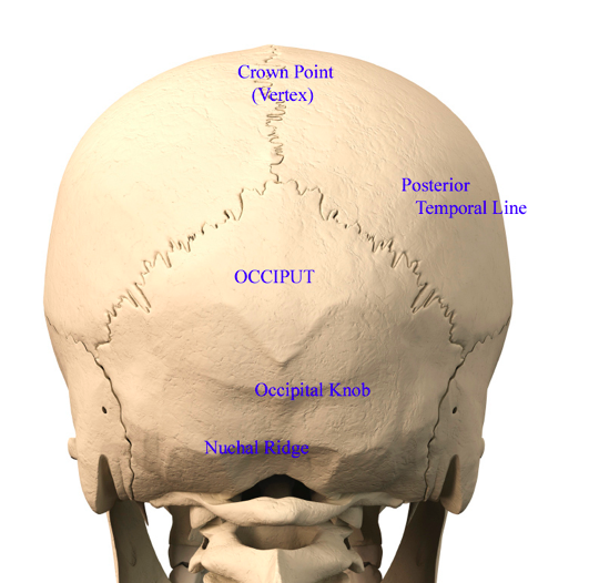


Back Of Head Skull Anatomy Dr Barry Eppley Indianapolis Explore Plastic Surgery
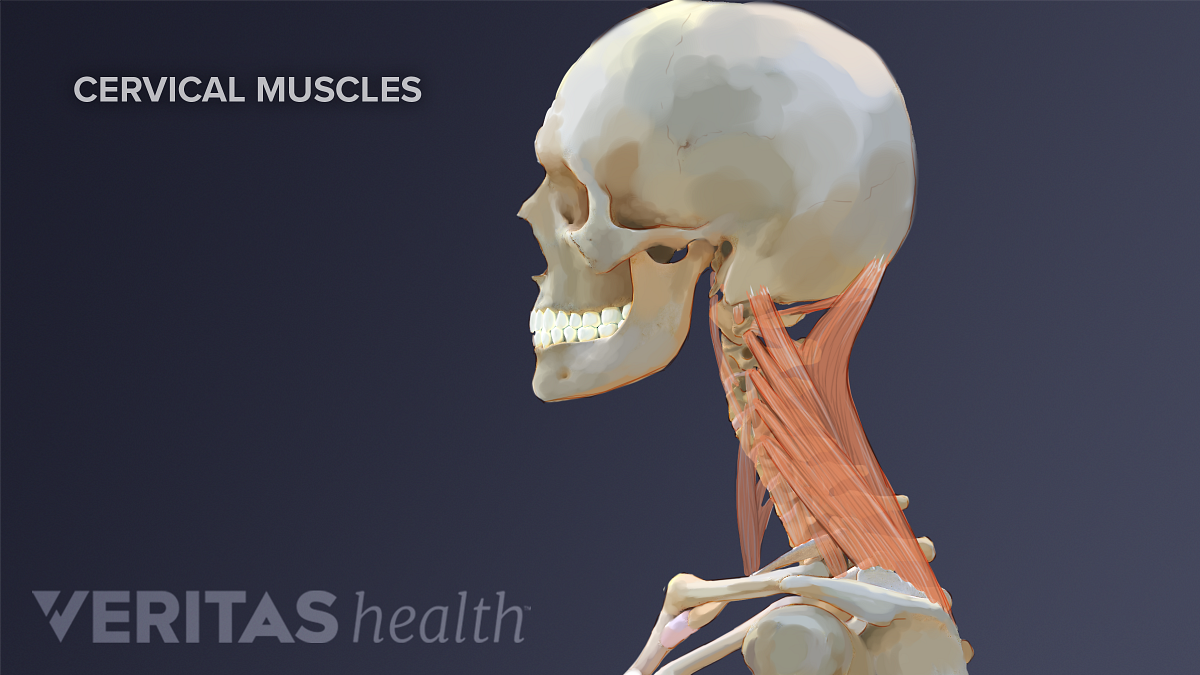


Neck Muscles And Other Soft Tissues


7 3 The Skull Anatomy Physiology


Skull Anatomy Terminology Dr Barry L Eppley


Skull Back View The Human Skull Is A Bony Structure The Head In The Skeleton Which Supports The Structures Of The Face And Canstock
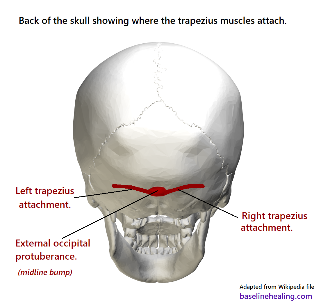


Upper Body To Base Line Connection The Trapezius Muscles
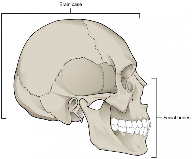


The Skull Anatomy And Physiology I


Skull Definition Anatomy Function Britannica


Why Do I Have A Bone Bump On The Back Of My Skull Quora
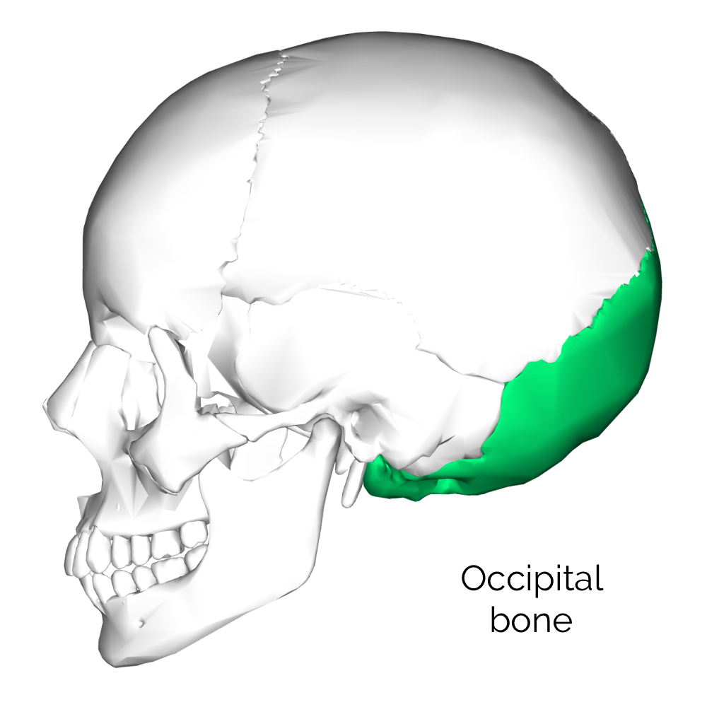


Bones Of The Skull Skull Osteology Anatomy Geeky Medics
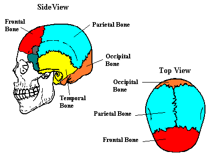


Neuroscience For Kids The Skull


No Teens Aren T Growing Skull Horns Because Of Cellphones Time


Anatomy Anatomy Massage Therapy Migraine


External Occipital Crest Canvas Prints Fine Art America


Skull Wikipedia


Bone Structure Of The Face An Overview Of Dental Anatomy Continuing Education Course Dentalcare Com


Sagittal Skull Surgery For Peaked Head Shape Dr Eppley
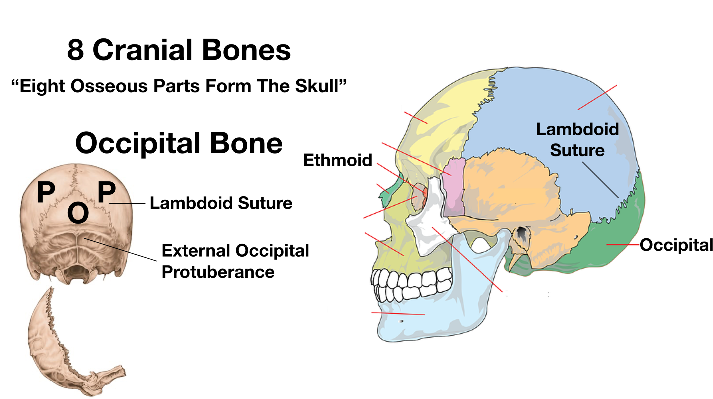


Skull Anatomy Cranial Bone And Suture Labeled Diagram Names Mnemonic Ezmed


Human Body Skull Anatomy External Occipital Protuberance Human Back Of Skull Face Human Png Pngegg


Human Head Skull And Cervical Vertebrae Rear View Stock Photo Download Image Now Istock


Skull Anatomy Terminology Dr Barry L Eppley
/male-skull-in-profile-with-transparent-head-on-white-background-1092338382-d031fe3a88fa4462b0ba082a1ec64302.jpg)


Occipital Bone Anatomy Function And Treatment
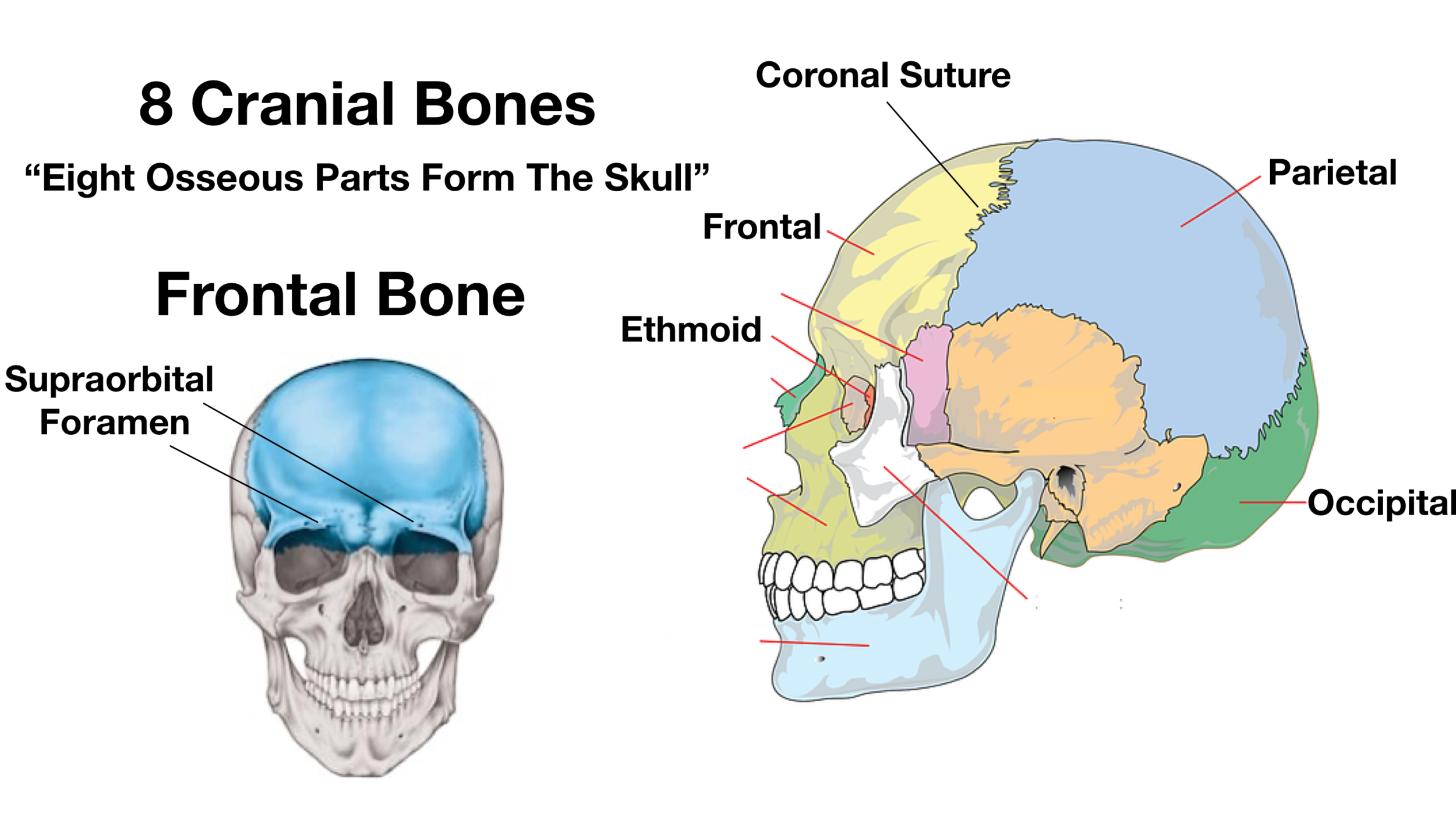


Skull Anatomy Cranial Bone And Suture Labeled Diagram Names Mnemonic Ezmed


Skull Anatomy Terminology Dr Barry L Eppley


Skull Scalp And Superficial Face


Skull Anatomy Terminology Dr Barry L Eppley
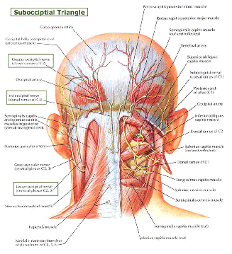


Occipital Nerve Blocks Complete Spine And Pain Care
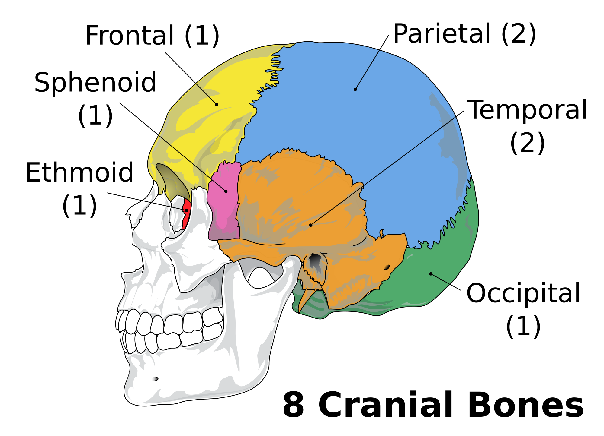


The Skull Boundless Anatomy And Physiology
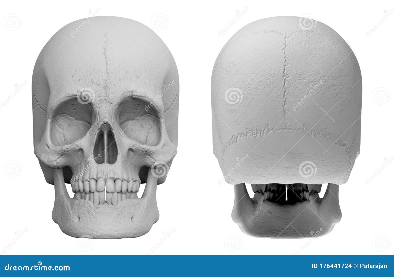


Side Back Human Skull Stock Illustrations 164 Side Back Human Skull Stock Illustrations Vectors Clipart Dreamstime


Bones Of The Head And Neck Skull And Cervical Spine Preview Human Anatomy Kenhub Youtube
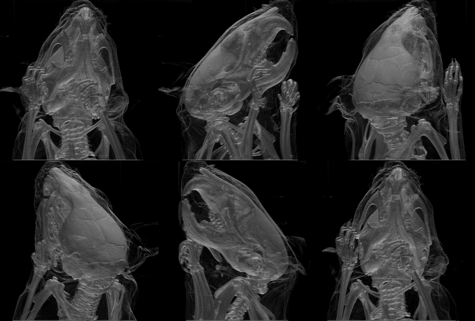


Skull Wikipedia


The Skull Anatomy And Physiology


Human Head Neck Skull Anatomy Medical Anatomical Chart Educationalmodel Com Amazon Com Books


Human Skull Back View Canvas Print Barewalls Posters Prints Bwc
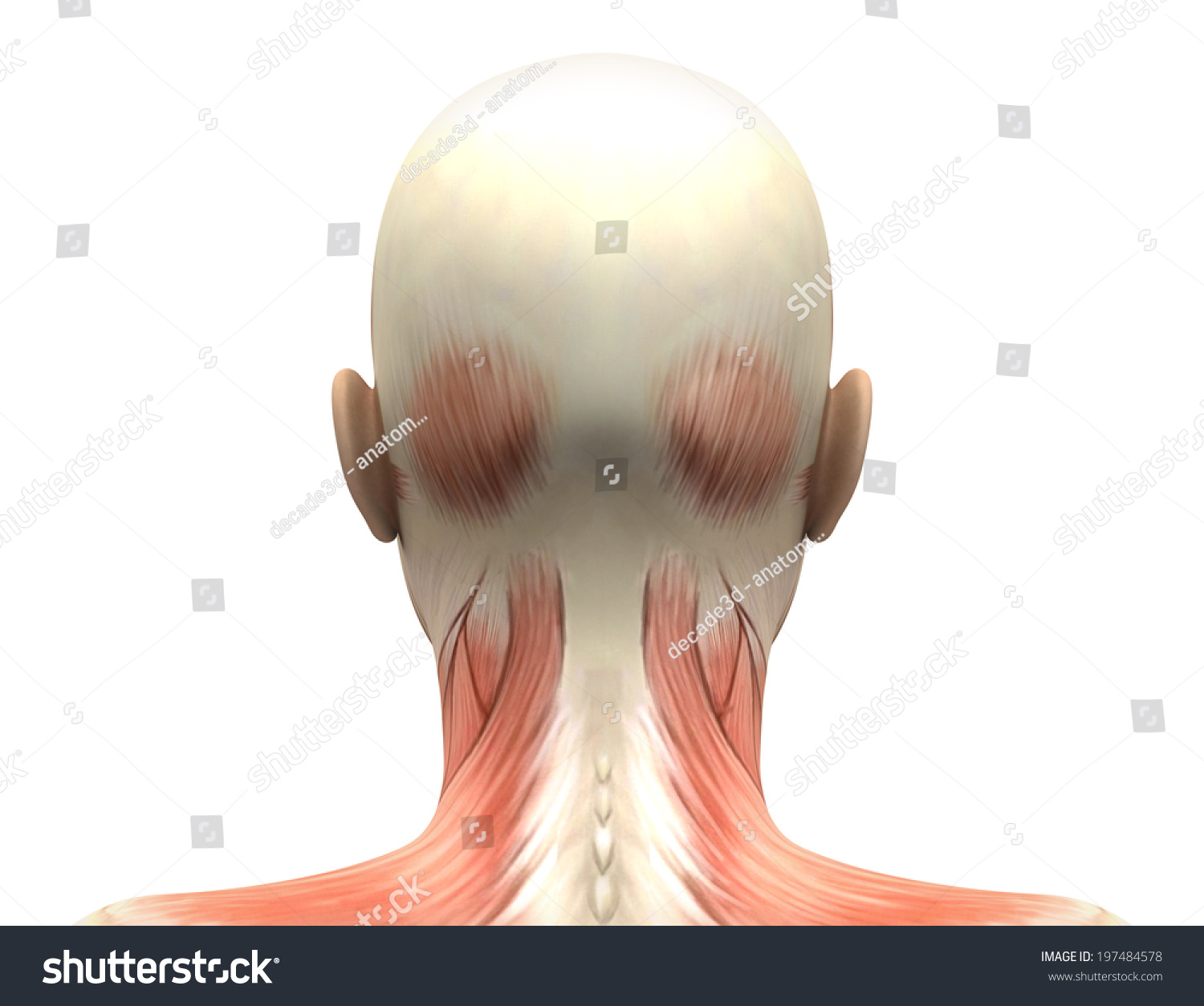


Female Head Muscles Anatomy Back View Stock Illustration


1
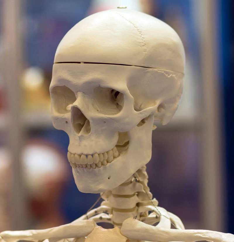


Temporal Bone Anatomical Diagram Function And Injuries


Very Detailed And Scientifically Correct Human Skull Back View On White Background Anatomy Image Stock Photo Picture And Royalty Free Image Image
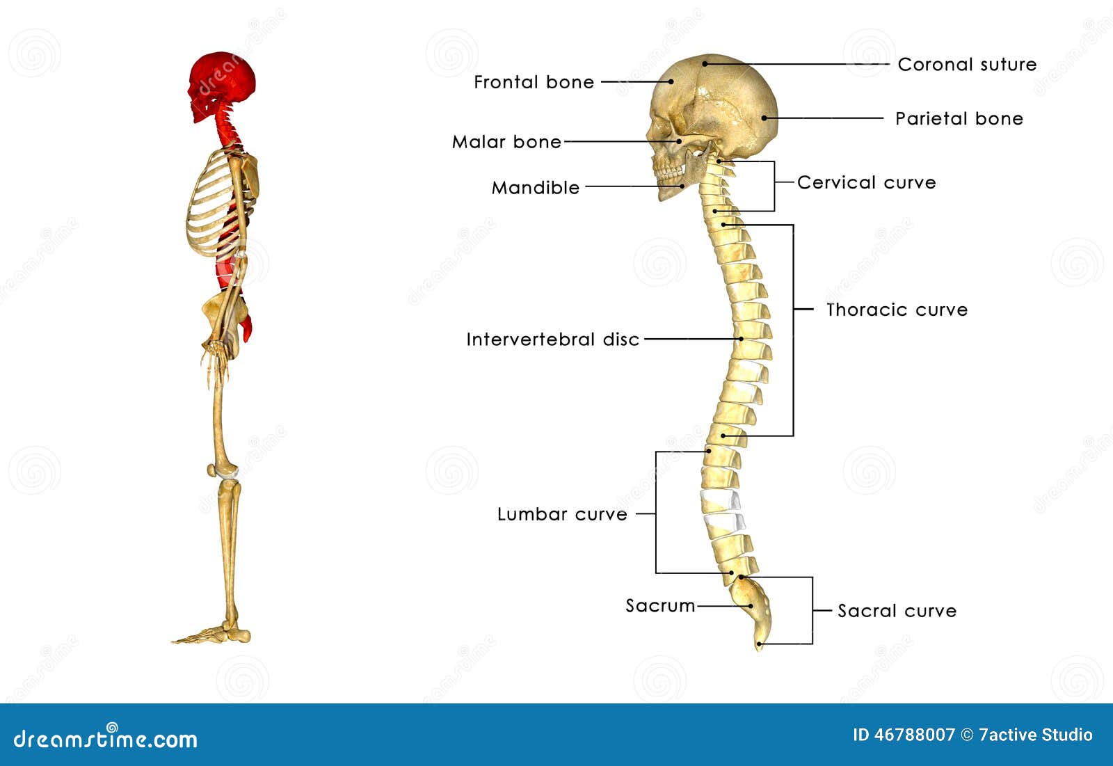


Back Bone With Skull Side View Stock Illustration Illustration Of Head Gland


Neck Bone Sticking Out


Anatomy Back Of Skull 2 Diagram Quizlet


3


Poor Posture Due To Smartphone Use Leads To Horn Bone Growth In Skull Youtube
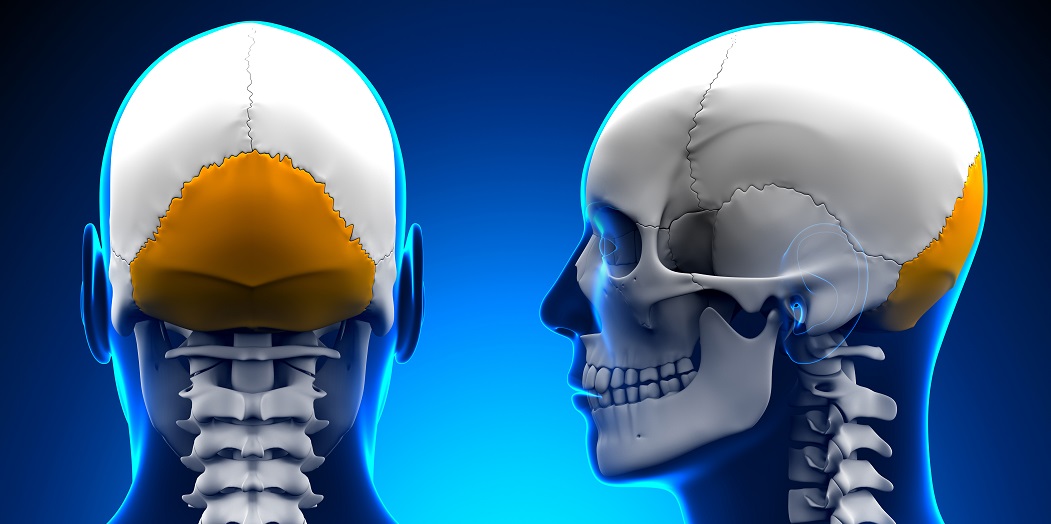


Occipital Bone The Definitive Guide Biology Dictionary
:background_color(FFFFFF):format(jpeg)/images/article/en/posterior-and-lateral-views-of-the-skull/U0Gu2npm5ZRSP1eZgc4Jbw_Posterior_view_of_skull.png)


Posterior And Lateral Views Of The Skull Anatomy Kenhub


Skull Back Images Stock Photos Vectors Shutterstock


Posterior And Lateral Views Of The Skull Anatomy Kenhub


Anatomy Of Back Of Head Anatomy Drawing Diagram


Human Skull 3 4 Back View Skull Reference Human Skull Skull Anatomy


Upper Cervical Spine Disorders Anatomy Of The Head And Upper Neck


A P Lab Bones Skull Anatomy Flashcards Quizlet
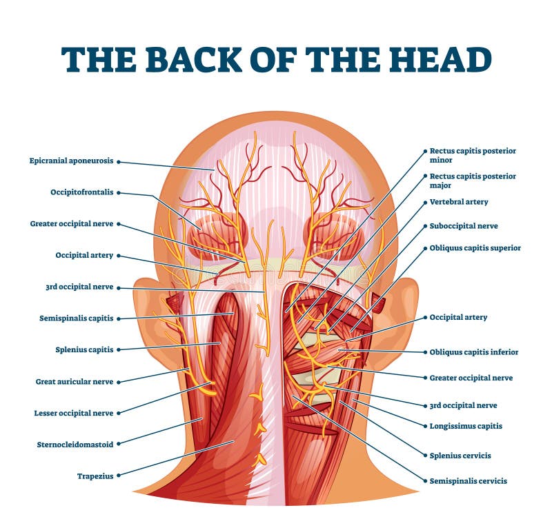


Back Of The Head Muscle Structure And Nerve System Diagram Stock Vector Illustration Of Labeled Muscle
:background_color(FFFFFF):format(jpeg)/images/library/9490/skull-posterior-lateral-views_english.jpg)


Posterior And Lateral Views Of The Skull Anatomy Kenhub


12 Types Of Bump On The Back Of The Head
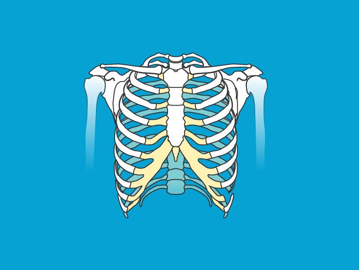


Skull Pictures Anatomy Diagram Body Maps


Bumps Ridges And Soft Spots On A Baby S Head When Should You Worry Quest For Health Kc
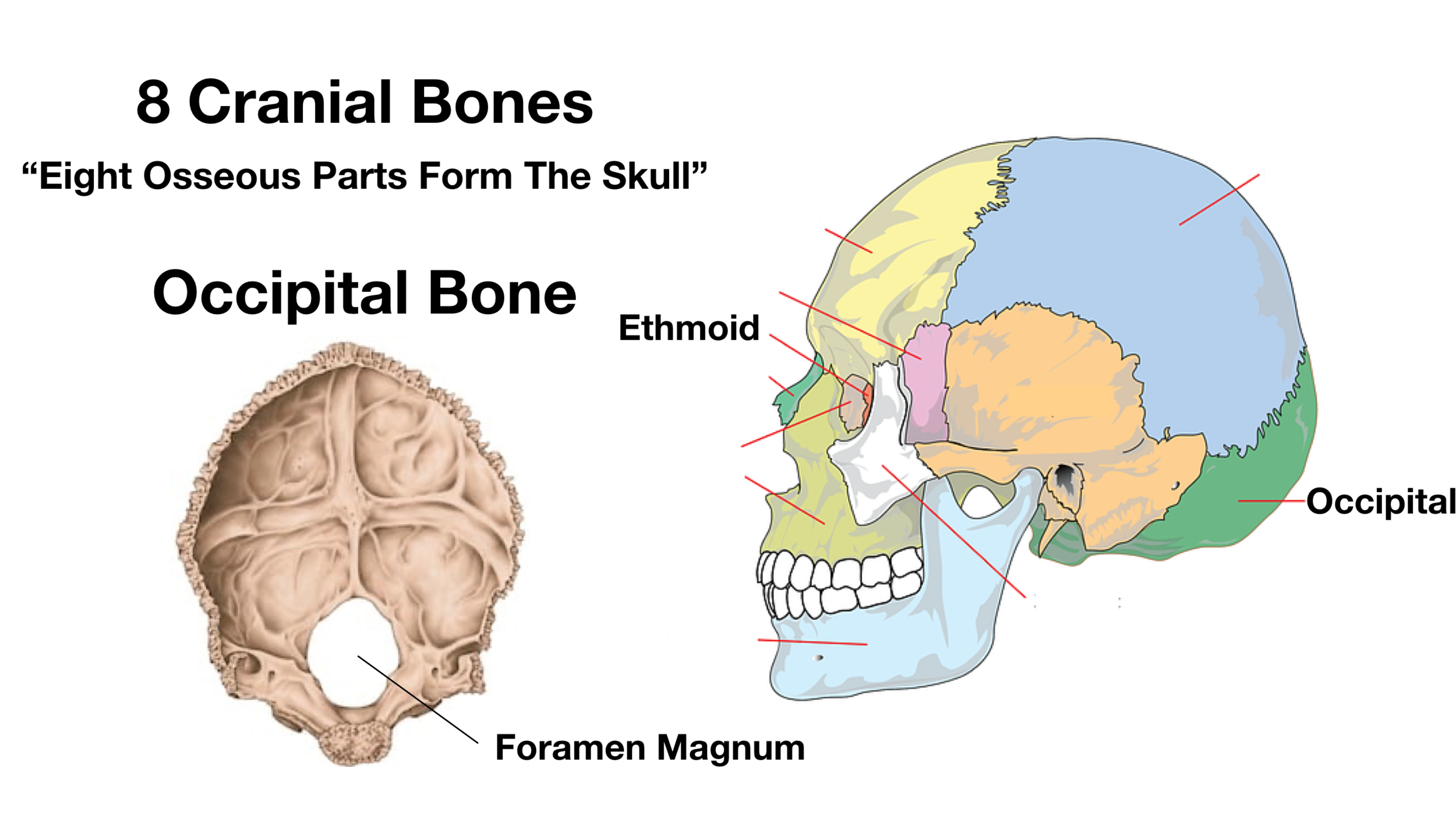


Skull Anatomy Cranial Bone And Suture Labeled Diagram Names Mnemonic Ezmed
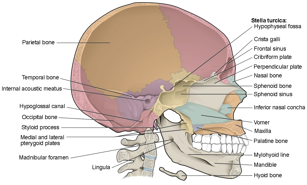


The Skull Anatomy And Physiology I


Flashcards Bones Axial Skeleton Skull Cavities Skull Anatomy Anatomy Bones Medical Anatomy
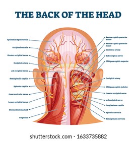


Occipital Images Stock Photos Vectors Shutterstock


Cervical Dysfunction And Pain In The Head And Neck Causes And Osteopathic Options
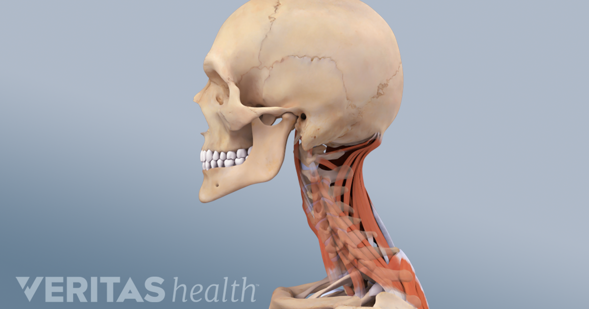


Cervicogenic Headache Causes And Risk Factors


Anatomy The Human Skull Youtube
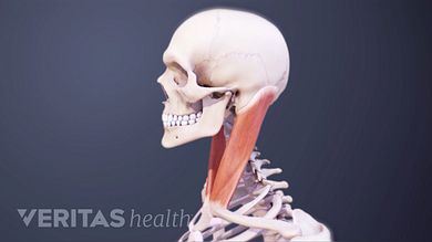


Neck Muscles And Other Soft Tissues
:max_bytes(150000):strip_icc()/human-skull-with-veins-and-arteries--rear-view--1174640349-490cb7f8593945c4b1690b152e6a4074.jpg)


Occipital Artery Anatomy Function And Significance


1


The Skull Boundless Anatomy And Physiology
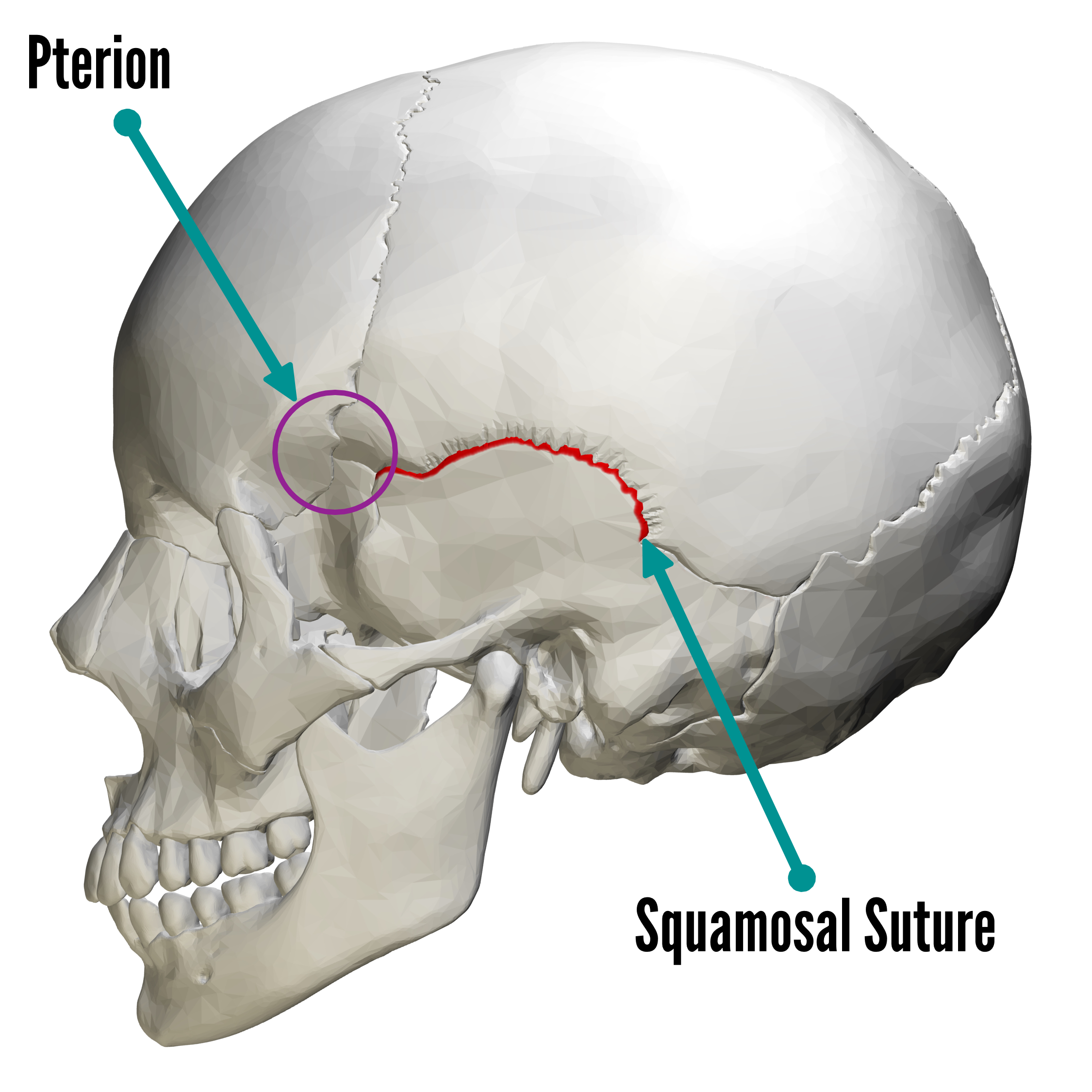


Bones Of The Skull Skull Osteology Anatomy Geeky Medics
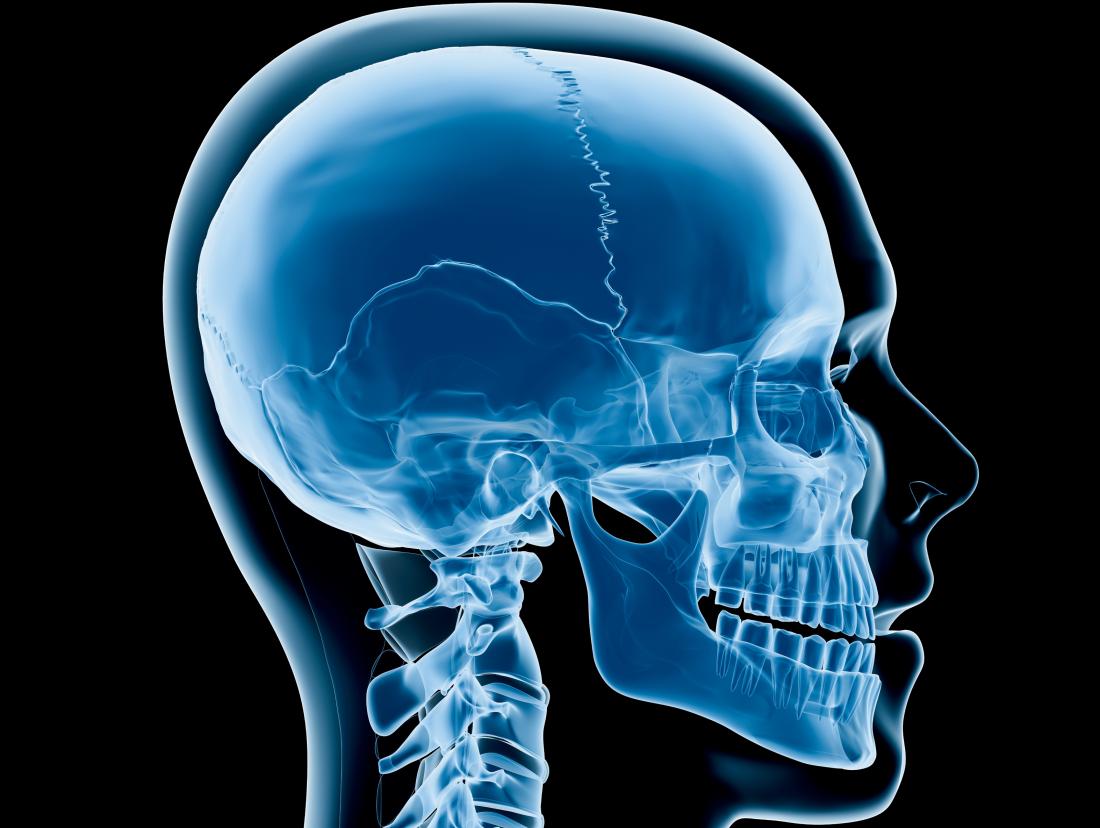


Craniosynostosis Types Causes Diagnosis And Treatment


Bones Of The Head Atlas Of Anatomy
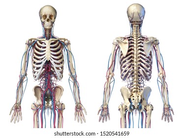


Skull Back Images Stock Photos Vectors Shutterstock


7 99 Aud 06 Anterior And Posterior Views Of Skull Anatomy Map 14 X25 Poster Ebay Collectibles Skull Anatomy Anatomy Bones Human Skull Anatomy


Lateral View Of Human Skull Anatomy By Alayna Guza Human Skull Anatomy Skull Anatomy Head Anatomy
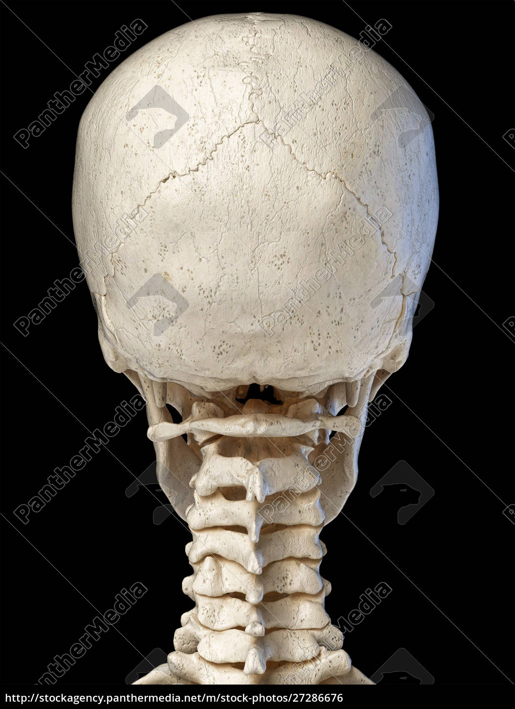


Human Skull Viewed From The Back Royalty Free Photo Panthermedia Stock Agency


7 3 The Skull Anatomy Physiology
:background_color(FFFFFF):format(jpeg)/images/library/10686/Posterior_view_of_the_skull.jpg)


Skull Anatomy Structure Bones Quizzes Kenhub
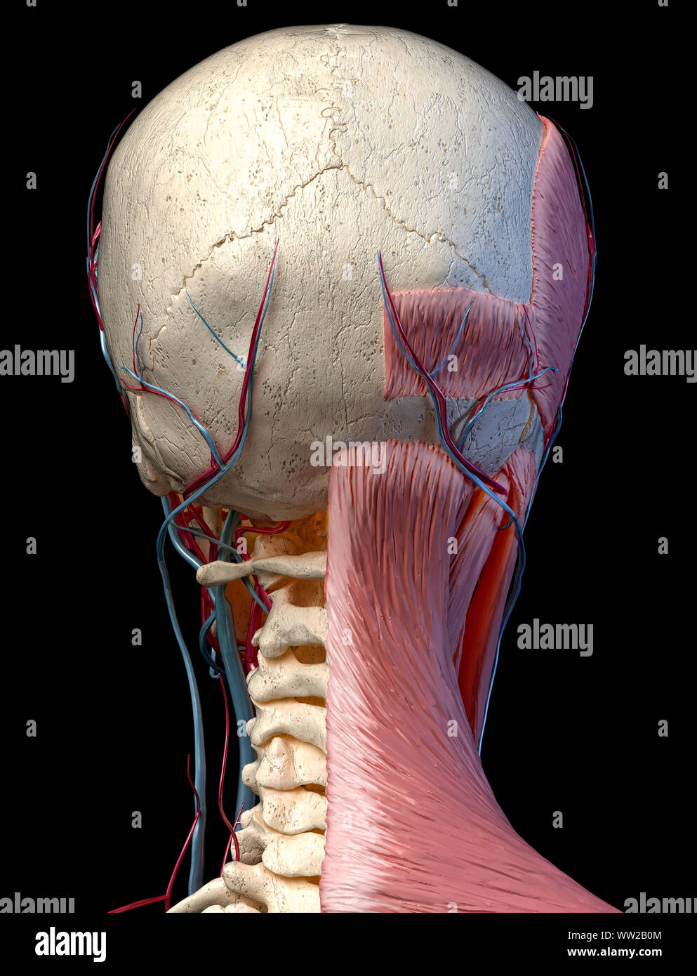


Human Anatomy 3d Illustration Of Head With Skull Blood Vessels And Muscles On Black Background Rear View Stock Photo Alamy
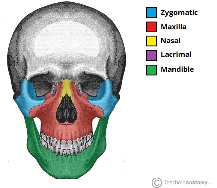


Bones Of The Skull Structure Fractures Teachmeanatomy
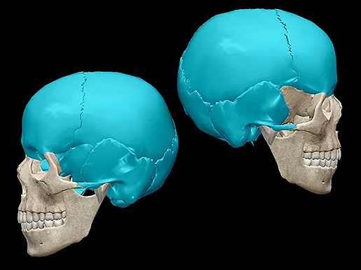


Five Fast Facts About Skull Anatomy
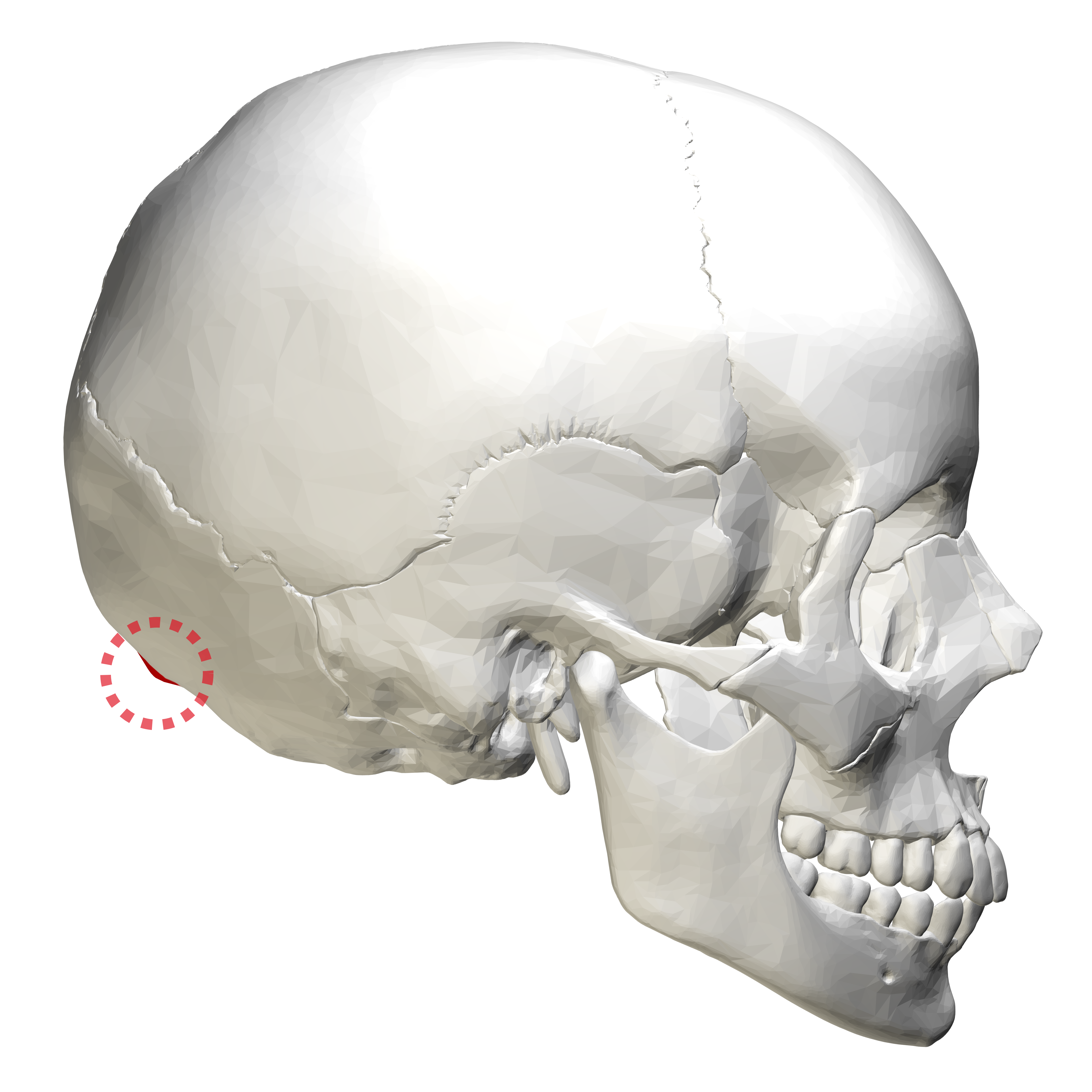


External Occipital Protuberance Wikipedia
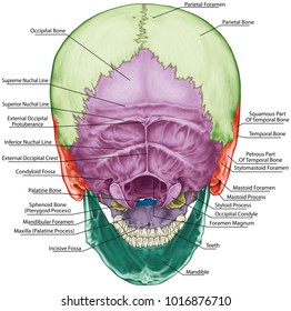


Skull Anatomy Images Stock Photos Vectors Shutterstock
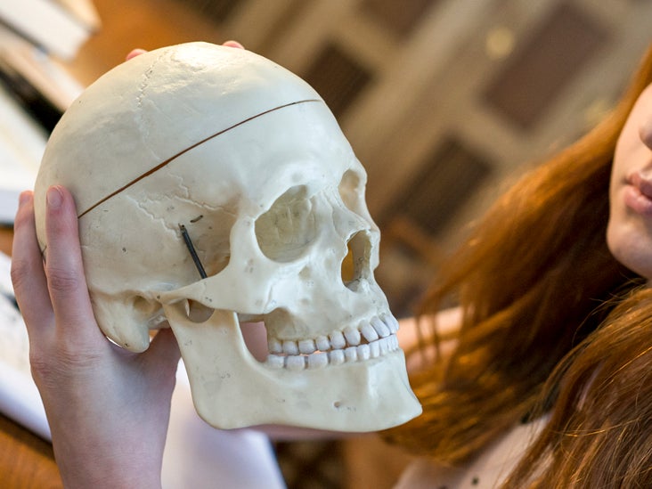


Cranial Bones Function And Anatomy Diagram Conditions Health Tips


The Skull Anatomy And Physiology I


Crown Of Head Conditions Injuries And More
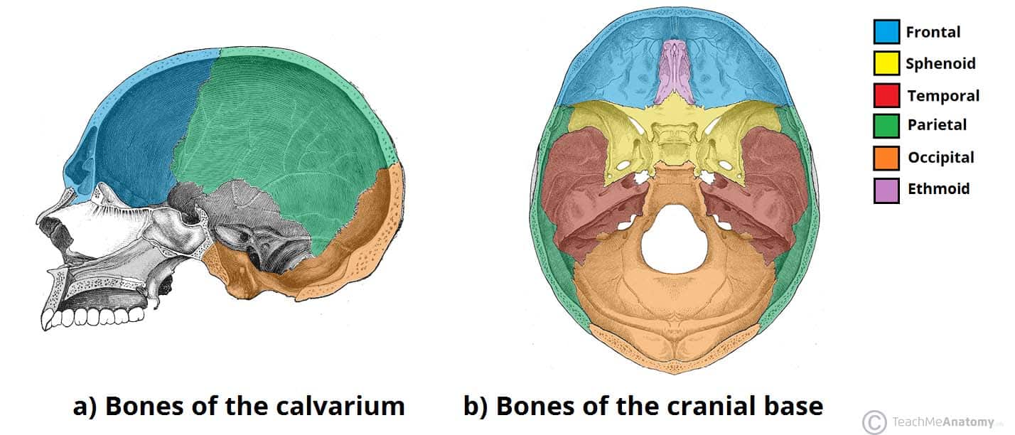


Bones Of The Skull Structure Fractures Teachmeanatomy


0 件のコメント:
コメントを投稿